


 598 692

targeted to the peripheral zone or throughout the whole
prostate. The number of injections per lobe varied from one
to three (total 2–6 injections). Preoperative imaging to
localize the SN was either with lymphoscintigraphy or
SPECT-CT, and when indocyanine green (ICG) was used,
preoperative imaging was omitted.
2.4.
Patient types
Men with PCa who were eligible for local treatment with
either prostatectomy or radiotherapy and at risk of nodal
metastases were included. If preoperative nodal imaging
was not performed, patients were staged as cN0.
2.5.
Type of outcome measures
The primary outcomes for diagnostic test accuracy (DTA)
were the nondiagnostic rate (NDR), sensitivity, specificity,
positive predictive value (PPV), negative predictive value
(NPV), and false positive (FP) and false negative (FN) rates,
all measured at patient level only. If DTA outcomes were not
reported in the original article, these were derived and
calculated from the available data. FN cases were defined as
patients with histologically negative SN whilst cancer was
found in other LNs in the PLND template
( Fig. 1, no. 4). FP
cases were defined as patients with SNs containing
metastases outside the (e)PLND template while the (e)PLND
template did not reveal any metastases
( Fig. 1, no. 7). Thus,
FP provides a measure of the additional diagnostic value of
SNB over and above PLND. For studies that reported
outcomes using alternative definitions of DTA elements,
the outcomes were recalculated and derived using the
above standardized definitions. Additional outcomes in-
cluded the proportion of histologically positive cases in SNB
only.
2.6.
Data analysis
Data from each study at patient level were summarized in
2 2 tables with SNB as the index test and (e)PLND as the
reference standard
( Fig. 1and Supplementary Table 1).
These tables were used to calculate sensitivity, specificity,
NPV, and PPV. Studies reporting insufficient data (eg,
studies reporting results only at the LN level and not the
patient level) were excluded. Owing to the expected
clinical heterogeneity in patient characteristics, defini-
tions, and thresholds and in types of intervention, a meta-
analysis was not planned; instead, a narrative synthesis
was carried out
[14] .All DTA outcomes are presented as
proportions (%) for individual studies and summarized as
median and interquartile range (IQR) for all studies
collectively. To explore the effect of heterogeneity on
the results, sensitivity analyses were planned for the
different definitions of SN, type of tracer, extent of PLND,
studies recruiting patients with intermediate- or high-risk
localized disease only, and studies with low to moderate
risk of bias (RoB). Additional analyses were performed on
histologically positive cases in SNB solely, and on patients
with LN metastases that were found outside the ePLND
template regardless of whether ePLND was positive or
negative.
2.7.
Assessment of RoB
To assess RoB, the RoB domains of the QUADAS-2 criteria
were used
[15] .RoB was scored as ‘‘low’’, ‘‘high’’, or
‘‘unclear’’ for the following domains: patient selection,
index test, reference standard, flow, and timing. RoB scoring
was performed independently by two reviewers (EW and
NG). Disagreement was resolved by discussion or with an
independent arbiter (HvdP or TBL).
3.
Evidence synthesis
3.1.
Quantity of evidence identified
The study selection process is outlined in the PRISMA flow
diagram
( Fig. 2 ). A total of 373 abstracts were screened,
including conference abstracts. No randomized controlled
trials were found. Fifty-four full-text publications were
retrieved for further screening, from which 21 studies met
the inclusion criteria
[16–36] .3.2.
Characteristics of the studies included
Data were included for 2509 patients from the 21 articles
included, of which 15 were prospective and six were
retrospective studies. Only one study
[16]was reported as a
conference abstract. SNB was performed in combination
with radical prostatectomy in 15 studies, with external
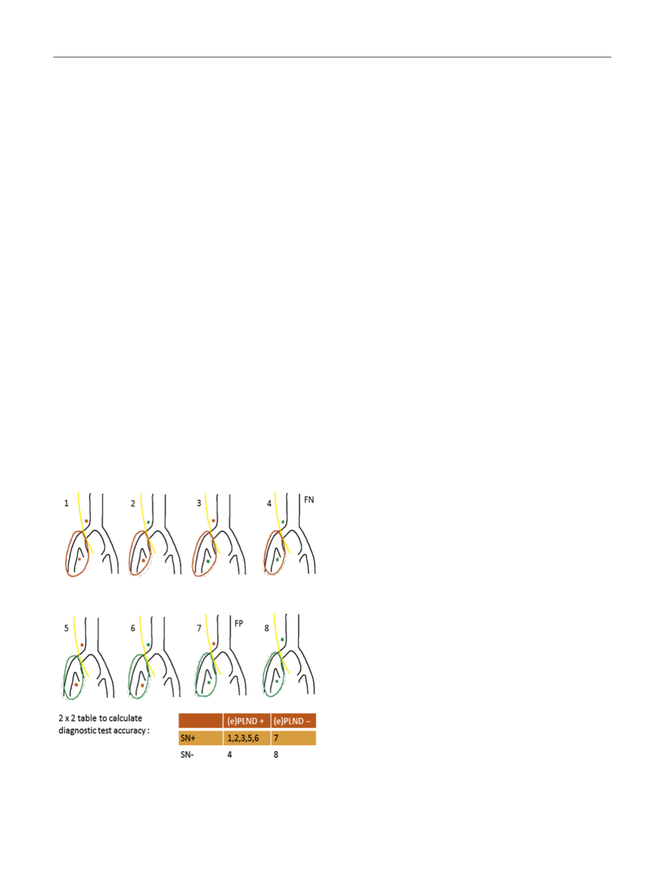
[(Fig._1)TD$FIG]
Fig. 1 – Variety of possible locations for sentinel nodes (SNs) and the
resulting variety for calculating false positive (FP) and false negative
(FN) results. No. 4 shows an FN case for which the SN was histologically
negative (green dot) while cancer was found in other LNs in the
dissection template (red oval). No. 7 shows an FP case defined as SN(s)
containing metastases outside the (e)PLND template (red dot) while the
(e)PLND template showed no metastases (green dot/oval).
E U R O P E A N U R O L O G Y 7 1 ( 2 0 1 7 ) 5 9 6 – 6 0 5
598